Understanding Your Vascular Lab Results: A Patient's Guide to ABI, Doppler, and Duplex Ultrasound

Blog Post
By South Valley Vascular
March 16, 2026

Understanding Your Vascular Lab Results: A Patient's Guide to ABI, Doppler, and Duplex Ultrasound

After a visit to a vascular specialist, you may leave with a set of numbers and terms from your diagnostic tests that can seem confusing. Understanding what these tests measure and what the results indicate is a key part of being an active participant in your own healthcare. The vascular lab is where we get a clear picture of what is happening inside your arteries and veins, and these non-invasive tests provide the crucial information we need to create your treatment plan.

At South Valley Vascular, we believe in patient education. This guide is designed to help you understand some of the most common tests performed in our accredited vascular laboratory, demystifying the results and empowering you to have more informed conversations with your doctor.

Ankle-Brachial Index (ABI): The Starting Point for PAD

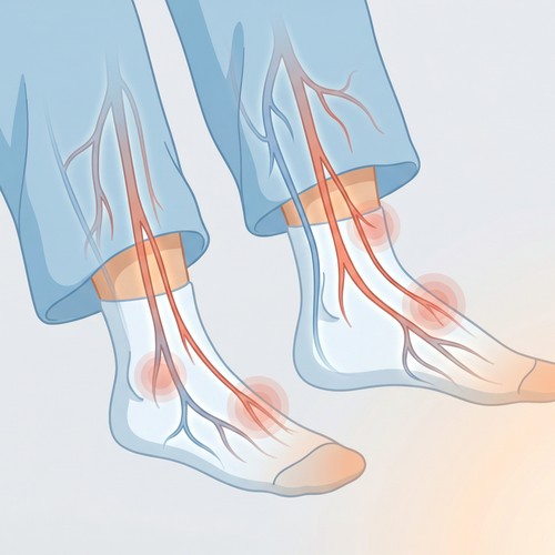
The Ankle-Brachial Index, or ABI, is a simple, quick, and painless test that is the first step in diagnosing Peripheral Artery Disease (PAD). It compares the blood pressure in your ankle to the blood pressure in your arm.

How it Works: A blood pressure cuff is placed on your arm and then on your ankle. A Doppler device, which uses sound waves to detect blood flow, is used to find your pulse. The systolic (top number) blood pressure is measured in both locations. The ankle pressure is then divided by the arm pressure to calculate the ABI.

What the Results Mean:

  • ABI of 1.0 to 1.4: This is the normal range. It indicates that you have healthy, unobstructed blood flow to your legs.
  • ABI of 0.9 to 0.99: This is considered borderline. It may indicate the very early stages of PAD, and your doctor will likely recommend risk factor management.
  • ABI of 0.5 to 0.89: This indicates mild to moderate PAD. Patients in this range often experience claudication (pain with walking).
  • ABI below 0.5: This suggests severe PAD. Patients in this range may have pain even at rest and are at a higher risk for developing non-healing ulcers or gangrene.
  • ABI above 1.4: This is also abnormal. It indicates that the arteries in your legs are stiff and calcified, which prevents them from being compressed by the blood pressure cuff. This is common in patients with diabetes or chronic kidney disease and also suggests the presence of significant vascular disease.

Doppler and Duplex Ultrasound: Visualizing Your Blood Flow

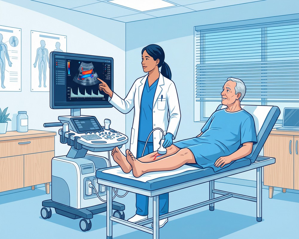
While the ABI provides a number, ultrasound tests provide a visual map of your circulatory system. These tests use high-frequency sound waves to create images of your blood vessels.

Doppler Ultrasound: This test is used to listen to the sound of blood moving through your vessels. The pitch and character of the sound can tell a trained technologist if blood flow is smooth and normal or turbulent and obstructed. It is a key part of the ABI test.

Duplex Ultrasound: This is the most common and comprehensive vascular lab test. It combines traditional ultrasound, which creates a black-and-white image of the vessel structure, with Doppler ultrasound, which adds color to show the direction and speed of blood flow. This "duplex" combination allows us to:

  • See the Anatomy: We can directly visualize the artery or vein, see the vessel walls, and identify any plaque buildup or blood clots.
  • Measure Blood Flow: The color imaging shows us exactly where blood is flowing and how fast it is moving. We can pinpoint the precise location of a blockage and measure its severity.

What We Look for During a Duplex Scan

When our registered vascular technologists perform a duplex scan, they are looking for specific signs of disease.

For Arterial Studies (PAD, Carotid Disease):

  • Plaque Buildup: We can see the plaque on the artery walls and measure how much it is narrowing the vessel (stenosis).
  • Velocity Changes: As an artery narrows, the blood flow speeds up as it squeezes through the blockage. We measure these velocity changes to grade the severity of the stenosis. A very high velocity indicates a significant blockage.
  • Waveform Analysis: The Doppler signal produces a waveform that has a characteristic shape in healthy arteries. In diseased arteries, this waveform becomes dampened and abnormal, which helps us identify problems.

For Venous Studies (DVT, Venous Insufficiency):

  • Compressibility: Healthy veins are easily compressed with light pressure from the ultrasound probe. A vein that will not compress is a classic sign of a blood clot (DVT).
  • Reflux (Leaky Valves): To test for venous insufficiency, we will squeeze your calf to push blood up the leg and then quickly release it. If the valves are working, the blood will not fall back down. On the ultrasound, we can see and measure the duration of any backward flow (reflux). Reflux lasting longer than 0.5 seconds is considered abnormal.

Your Partner in Understanding Your Health

Your vascular lab results are a vital piece of your health puzzle. They provide the objective data that, combined with your symptoms and physical exam, allows your vascular specialist to make an accurate diagnosis and recommend the most effective treatment.

Never hesitate to ask questions about your results. Understanding your condition is the first step toward managing it effectively. The team at South Valley Vascular is committed to ensuring you have a clear understanding of your diagnosis and your treatment options. If you have questions about your vascular health or need to schedule a diagnostic evaluation, please call us at (559) 625-4118.


Disclaimer: The information provided in this article is for educational purposes only and does not constitute medical advice. It is not intended to be a substitute for professional medical advice, diagnosis, or treatment. Always seek the advice of your physician or other qualified health provider with any questions you may have regarding a medical condition. Never disregard professional medical advice or delay in seeking it because of something you have read in this article.

{"@context": "https://schema.org", "@type": "MedicalWebPage", "name": "Understanding Your Vascular Lab Results: A Patient's Guide to ABI, Doppler, and Duplex Ultrasound", "description": "Confused by your vascular lab results? This patient's guide explains ABI, Doppler, and duplex ultrasound tests. Learn what your results mean from the experts at South Valley Vascular.", "url": "https://www.southvalleyvascular.com/blog/understanding-your-vascular-lab-results-a-patients-guide-to-abi-doppler-and-dupl", "datePublished": "2026-03-16", "publisher": {"@type": "MedicalOrganization", "name": "South Valley Vascular", "url": "https://www.southvalleyvascular.com", "telephone": "(559) 625-4118", "address": {"@type": "PostalAddress", "addressLocality": "Visalia", "addressRegion": "CA", "addressCountry": "US"}}, "medicalAudience": {"@type": "MedicalAudience", "audienceType": "Patient"}}