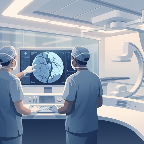
When it comes to diagnosing vascular conditions, physicians need to see inside the body's complex network of blood vessels without invasive procedures. This is where vascular ultrasound technology shines, offering detailed, real-time images of blood flow and vessel structure without radiation, contrast dyes, or surgical intervention.
Vascular ultrasound has revolutionized the diagnosis and management of circulatory conditions, allowing specialists to detect problems earlier, monitor disease progression more accurately, and plan interventions with greater precision. At South Valley Vascular, this technology forms the cornerstone of our diagnostic approach, providing our patients throughout the Central Valley with access to state-of-the-art vascular assessment.
Vascular ultrasound utilizes high-frequency sound waves—well beyond the range of human hearing—to create detailed images of blood vessels and blood flow. The basic principles are elegantly simple:
Sound Wave Transmission: A handheld device called a transducer emits sound waves that travel through the skin and other tissues.
Tissue Interaction: These sound waves bounce off blood cells and vessel walls, creating echoes that return to the transducer.
Signal Processing: The ultrasound machine processes these echoes, converting them into visual images displayed on a monitor.
Doppler Effect: By measuring changes in the frequency of returned sound waves, the system can determine the direction and speed of blood flow—a technique known as Doppler ultrasound.
What makes this technology particularly valuable is its ability to provide both structural and functional information simultaneously. Not only can vascular specialists see the physical condition of your blood vessels, but they can also assess how effectively blood is flowing through them.
At South Valley Vascular, we perform several specialized types of vascular ultrasound, each designed to evaluate specific aspects of circulatory health:
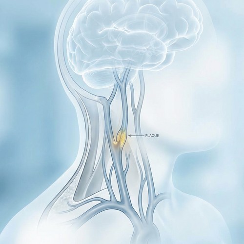
This examination focuses on the carotid arteries in your neck, which supply blood to your brain. The test can:
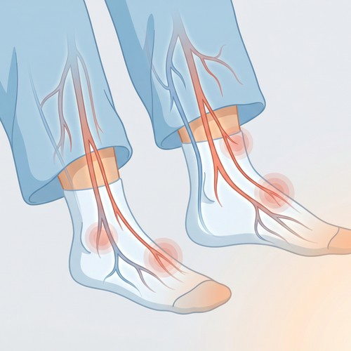
This assessment examines the arteries in your extremities, particularly the legs, to:
Focusing on the veins, particularly in the legs, this examination can:
This specialized examination looks at major blood vessels in the abdomen to:
For patients with dialysis access sites, ultrasound can:
Many patients express anxiety about medical tests, often due to uncertainty about what to expect. At South Valley Vascular, we prioritize patient comfort and understanding throughout the diagnostic process.
Preparation Requirements: Most vascular ultrasounds require minimal preparation. For abdominal studies, you may be asked to fast for 8-12 hours before the examination to reduce intestinal gas that could obscure imaging. For other studies, no special preparation is typically needed.
Medication Considerations: Generally, you should continue taking your regular medications unless specifically instructed otherwise.
Clothing Recommendations: Wear comfortable, loose-fitting clothing that can be easily adjusted to expose the area being examined. For lower extremity studies, consider wearing shorts; for upper extremity studies, a short-sleeved shirt is ideal.
Initial Assessment: Your technologist will begin by reviewing your medical history and the specific reason for your examination.
Positioning: You'll be positioned comfortably on an examination table, with the area being studied exposed.
Ultrasound Gel Application: A water-based gel will be applied to your skin. This gel may feel cool initially but ensures good contact between the transducer and your skin, allowing sound waves to transmit effectively.
The Scanning Process: The technologist will press the transducer against your skin and move it slowly over the area being examined. For Doppler studies, you may hear pulse-like sounds as the equipment detects blood flow.
Patient Participation: You may be asked to adjust your position slightly or to hold your breath briefly during certain parts of the examination.
Duration: Most vascular ultrasound examinations take between 30-60 minutes, depending on the complexity and extent of the study.
Immediate Activities: Once the examination is complete, the gel will be wiped off your skin, and you can resume normal activities immediately.
Results Timeline: At South Valley Vascular, a board-certified vascular specialist interprets all ultrasound studies. In many cases, preliminary results can be discussed immediately after the examination, with full results available within 1-2 business days.
Follow-up Planning: Based on your results, our team will recommend appropriate next steps, which may include treatment options, additional testing, or monitoring protocols.
South Valley Vascular has invested in cutting-edge ultrasound technology that goes beyond basic vascular assessment:
Traditional ultrasound provides two-dimensional images, but our advanced systems can generate three-dimensional reconstructions of blood vessels, with the fourth dimension being time (showing movement). This technology offers:
For certain complex cases, we may utilize contrast-enhanced ultrasound, which involves:
Our portable ultrasound capabilities allow for:
Vascular ultrasound offers several significant advantages over other imaging modalities:
Non-Invasive Nature: Unlike angiography, which requires arterial puncture and contrast injection, ultrasound is completely external and non-invasive.
Radiation-Free: Unlike CT scans or X-rays, ultrasound uses no ionizing radiation, making it safe for repeated use and for vulnerable populations like pregnant women.
Real-Time Imaging: Ultrasound provides dynamic, real-time images of blood flow, offering functional information that static imaging cannot.
Cost-Effectiveness: Vascular ultrasound is generally less expensive than CT angiography, MR angiography, or invasive angiography.
Accessibility: With multiple locations throughout the Central Valley, South Valley Vascular makes this advanced technology accessible to communities that might otherwise face geographical barriers to specialized care.
Vascular ultrasound plays a crucial role in diagnosing numerous circulatory conditions:
Ultrasound can detect and quantify narrowing in the carotid arteries, helping to assess stroke risk and determine whether interventions like carotid endarterectomy or stenting might be beneficial.
Through arterial duplex studies, ultrasound can identify locations and severity of arterial narrowing or blockages in the legs, guiding treatment decisions from lifestyle modifications to interventional procedures.
Venous ultrasound can demonstrate valve dysfunction in leg veins, helping to diagnose chronic venous insufficiency and guide treatments ranging from compression therapy to minimally invasive vein procedures.
Ultrasound is the gold standard for diagnosing blood clots in the deep veins, allowing for prompt treatment to prevent complications like pulmonary embolism.
Regular ultrasound monitoring allows for precise measurement of aneurysm size and growth rate, informing decisions about when surgical intervention becomes necessary.
For patients requiring dialysis, ultrasound can quickly identify issues with fistulas or grafts, allowing for timely intervention to maintain access function.
At South Valley Vascular, ultrasound technology extends beyond initial diagnosis to guide treatment decisions and monitor outcomes:
Procedural Planning: Detailed ultrasound mapping helps our specialists plan the most effective and least invasive approach for procedures like varicose vein treatment or arterial interventions.
Intraoperative Guidance: During certain procedures, ultrasound provides real-time visualization, enhancing precision and safety.
Post-Procedure Assessment: Immediate post-procedure ultrasound confirms technical success and establishes a baseline for future comparison.
Long-Term Monitoring: Sequential ultrasound examinations allow for objective assessment of treatment effectiveness and disease progression over time.
Many patients have questions about coverage for diagnostic testing:
Medicare Coverage: Medicare typically covers vascular ultrasound when medically necessary for diagnosing or monitoring vascular conditions.
Private Insurance: Most private insurance plans cover vascular ultrasound studies when ordered for appropriate clinical indications. Our office staff can help verify your specific coverage.
Screening Studies: Coverage for screening (in asymptomatic individuals) varies by insurance plan and risk factors. For example, Medicare covers one-time abdominal aortic aneurysm screening for eligible beneficiaries.
Self-Pay Options: For patients without coverage for specific studies, South Valley Vascular offers reasonable self-pay rates and payment plans.
To ensure the most accurate results from your vascular ultrasound examination:
Follow Preparation Instructions: Adhere to any fasting or medication instructions provided by our office.
Stay Hydrated: Unless instructed to fast, maintain good hydration before your appointment, as this improves vascular visualization.
Bring Information: Bring a list of current medications, relevant medical records, and previous vascular study results if performed elsewhere.
Wear Appropriate Clothing: Choose clothing that allows easy access to the area being examined.
Allow Sufficient Time: Plan to arrive 15 minutes before your appointment to complete any necessary paperwork.
The quality of vascular ultrasound depends not only on advanced equipment but also on the expertise of those performing and interpreting the studies. At South Valley Vascular, all ultrasound examinations are performed by registered vascular technologists and interpreted by board-certified vascular specialists.
This combination of cutting-edge technology and specialized expertise ensures that patients throughout the Central Valley receive accurate diagnoses, appropriate treatment recommendations, and ongoing monitoring of their vascular health.
If you're experiencing symptoms that might indicate a vascular condition, or if you have risk factors that warrant screening, contact South Valley Vascular today to schedule a consultation. With locations in Visalia, Hanford, Porterville, and Fresno, expert vascular ultrasound is never far away.
This article is provided for informational purposes only and does not constitute medical advice. Always consult with a qualified healthcare provider for diagnosis and treatment of medical conditions.