Vascular issues are far from an uncommon issue for Americans or the rest of the world. According to UCSF Health, cardiovascular disease is the number one cause of death in the United States for both men and women. In fact, 950,000 people die from cardiovascular disease each year. This displays why it is so important to manage cardiovascular health and discover these conditions as early as possible. Proper finding and diagnosis of these conditions offer the best chance for treatment and improvement. One of the most important tools in examining the arteries and veins is ultrasound technology.
Continue below to learn more about ultrasounds and how they are used in diagnosing and treating vascular issues.
Ultrasound technology uses sound to form an image of structures within the body. A technician will use an instrument called a transducer that sends sound waves into the body. These sound waves contact tissues and organs within the body and bounce back to the instrument, which is used to form an image that will be displayed on a monitor.
There are a few different types of ultrasound used for vascular issues.
This type of ultrasound is used for those with varicose veins. The technician will use special techniques to evaluate the valves within the veins of the legs. Abnormal valves can cause blood to pool within the legs, which is also known as venous insufficiency. Varicose veins or blood clots most commonly cause this condition.
Valves found to be functioning as they should are called competent valves, and those working incorrectly are called abnormal.
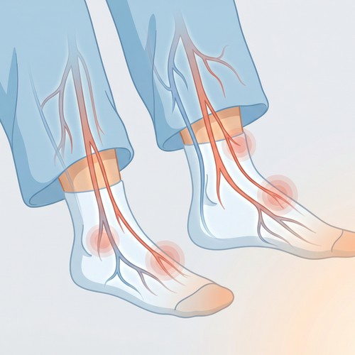
For those with Peripheral Artery Disease (PAD), a lower extremity arterial duplex ultrasound will likely be used. PAD is caused by a lack of blood flow to the legs, and this ultrasound method is used to evaluate any narrowing or plaque buildup in the leg arteries.
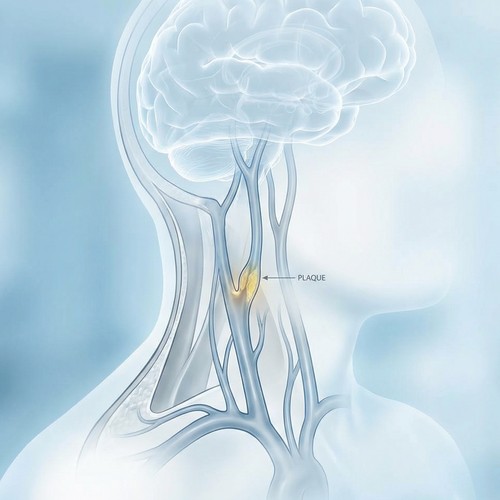
A carotid ultrasound examines the level of blood flow through the carotid arteries, which run along each side of the neck. These arteries supply blood from the heart up to the brain. It is also used to evaluate the thickness of the artery walls and check for the presence of clots.
A renal artery ultrasound checks the blood flow of the arteries that supply blood to the kidneys. This type will be used to look for blockages or narrowing or to monitor existing renal artery disease.
The above are not the only types of ultrasound that may be ordered by a patient's doctor. It will depend on the person's particular case, symptoms, and difficulties.
Some people may have heard the term sonogram used as well, which may make them wonder what's better, sonogram vs ultrasound? However, these are not actually two separate types of scans. They are sometimes used interchangeably, but they are not the same thing either.
Sonography just means using ultrasound technology for diagnostic reasons. Essentially, an ultrasound is the actual procedure, while a sonogram is the image produced by the procedure.
Are you struggling with a vascular condition or experiencing vascular-related symptoms? Then, South Valley Vascular can help get you the answers and treatment that you need.
We are part of the BASS Medical Group, which has helped tens of thousands of people with their vein-related conditions, and we can do the same for you. If you have any questions or would like to become a patient, call us at 559-625-4118 or head over to our contact page.