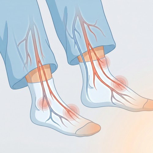
An abdominal aortic aneurysm is typically found in older men, especially those who are smokers. These are balloon-like bulges in the aorta, which is the largest artery and is responsible for carrying blood from the heart through the chest and torso. When the force of blood pumping is too much it will end up splitting the layers of the artery’s walls.
These aneurysms tend to grow slowly, typically without any symptoms whatsoever. However, some people do mention that they feel a pulsating feeling near their navel. Others mention feeling pain in their back, belly, or side. Oftentimes this is a sign that the aneurysm is about to rupture.
Symptoms of a leaking aneurysm include:
Symptoms of a ruptured aneurysm include:
Many times, the abdominal aortic aneurysm will be diagnosed for the first time during a chest X-ray, CT scan, or ultrasound for some unrelated reason. Anyone who has had abdominal aortic aneurysm symptoms will need an abdominal aortic aneurysm screening. This usually starts with a physical exam but doctors also use other tests, including:
Small aneurysms (under 5.5 cm), also known as slow-growing aneurysms, are not as likely to rupture as larger ones or ones that grow at a faster rate. Most doctors will simply monitor these with routine abdominal ultrasound instead of treating them.
Large aneurysms (over 5.5 cm), also known as fast-growing aneurysms, are the ones that are of more concern because of the greater likelihood that they will rupture. Such ruptures can be life-threatening because they can result in serious complications such as internal bleeding. The larger the aneurysm is, the more likely it will need to be treated with surgery, especially if it is leaking blood or causing other symptoms.
If you need an abdominal aortic aneurysm screening and live near the Southern San Joaquin Valley, call (559) 625-4118 or visit South Valley Vascular today. The healthcare professionals here have many years of experience, so you can trust them to take great care of you.